


.png)
HSG – HysterosalpingoGraphy
Hysterosalpingography (HSG) is a specialized radiographic technique used to examine the inside of the uterus and fallopian tubes. It involves injecting a contrast dye into the uterine cavity and taking X-ray images to visualize the reproductive structures. This procedure is mainly used in the evaluation of female infertility, as it can identify abnormalities or blockages that may be preventing conception. HSG provides detailed images that help doctors diagnose issues related to the uterine shape, size, and the patency of the fallopian tubes.
The primary indication for HSG is the assessment of infertility, especially when a woman has been unable to conceive after one year of unprotected intercourse, or six months if the woman is over 35. It helps detect uterine abnormalities such as congenital malformations, polyps, fibroids, and intrauterine adhesions, as well as blockages or damage in the fallopian tubes. By identifying these issues, healthcare providers can better understand the underlying causes of infertility and recommend appropriate treatments, such as surgical intervention or assisted reproductive technologies like in vitro fertilization (IVF).
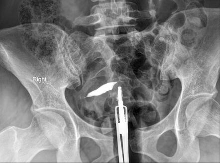
During the HSG procedure, the patient lies on an X-ray table, and a speculum is inserted into the vagina to visualize the cervix. The cervix is then cleaned, and a thin catheter is gently inserted through the cervical canal into the uterine cavity. A contrast dye is slowly injected through the catheter, filling the uterine cavity and fallopian tubes. As the dye moves through these structures, a series of X-ray images are taken to capture its flow. This process allows the radiologist to observe the shape and condition of the uterus and the openness of the fallopian tubes in real time.
The results of an HSG can reveal various conditions that affect fertility. Normal results show a uniformly shaped uterine cavity and free passage of dye through open fallopian tubes, indicating no blockages. Abnormal findings may include uterine anomalies like a septate or bicornuate uterus, polyps, fibroids, or scarring (Asherman's syndrome). Tubal blockages, often caused by pelvic inflammatory disease (PID), endometriosis, or previous surgeries, can also be identified. The identification of such abnormalities helps guide further diagnostic and therapeutic steps, improving the chances of successful conception.
HSG is a valuable diagnostic tool with several benefits, including its ability to provide detailed images of the reproductive organs non-invasively. However, the procedure is not without risks. Some patients may experience discomfort, cramping, or mild pain during and after the procedure. There is also a small risk of infection or allergic reaction to the contrast dye. Despite these risks, HSG remains a critical procedure in the diagnosis and management of infertility, offering essential insights into the health and functionality of the female reproductive system.