


.png)
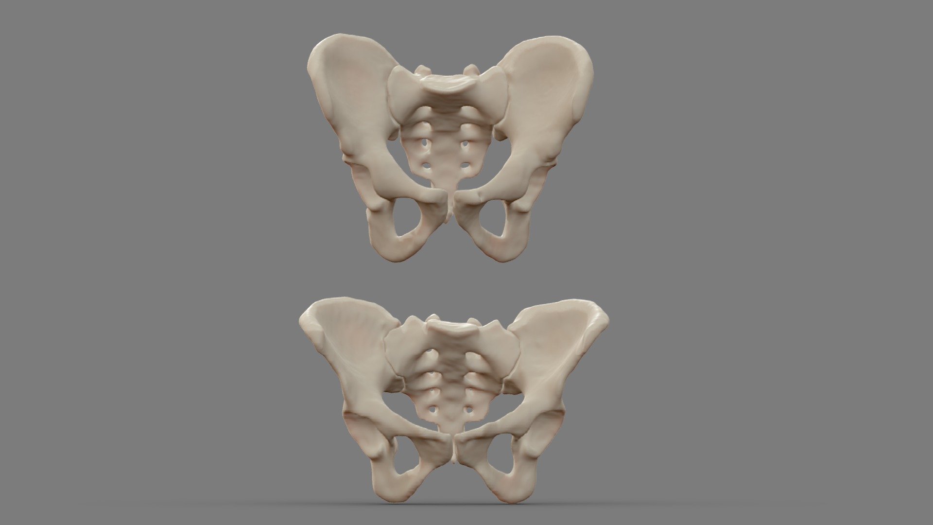
Pelvic 3D Scan
A pelvic 3D scan is a sophisticated medical imaging technique that provides a detailed three-dimensional view of the pelvis, a crucial structure in the human body. The pelvis supports the spine and houses several vital organs, including parts of the digestive and urinary systems. Utilizing advanced technologies such as CT (computed tomography) or MRI (magnetic resonance imaging), a pelvic 3D scan can capture comprehensive images, allowing healthcare professionals to examine the pelvis with greater accuracy and detail than traditional two-dimensional scans.
The process of obtaining a pelvic 3D scan typically involves the patient lying on a table that slides into the scanning machine. For a CT scan, X-rays are used to capture multiple cross-sectional images of the pelvic area, which are then combined by a computer to create a 3D model. In the case of an MRI, powerful magnets and radio waves generate detailed images of the soft tissues. The resulting 3D images provide a holistic view of the pelvic anatomy, highlighting bones, muscles, organs, and blood vessels.
One of the primary benefits of a pelvic 3D scan is its ability to aid in the diagnosis and treatment planning of various medical conditions. For instance, it can help detect fractures, tumors, infections, and congenital abnormalities. Additionally, the detailed imagery allows for precise surgical planning, improving the outcomes of procedures such as hip replacements or tumor removals. By offering a clear and comprehensive view of the pelvic region, these scans can significantly enhance the accuracy of medical interventions.
Furthermore, pelvic 3D scans are invaluable in monitoring the progression of diseases and evaluating the effectiveness of treatments. For patients with chronic conditions such as arthritis or cancer, regular 3D scans can track changes in the pelvic anatomy over time. This ongoing monitoring enables healthcare providers to adjust treatment plans promptly, ensuring the best possible care. Moreover, the non-invasive nature of the scans makes them a preferred option for repeated imaging, minimizing discomfort and risk for patients.
In conclusion, pelvic 3D scans represent a critical advancement in medical imaging, offering detailed and accurate views of the pelvic region. These scans facilitate precise diagnosis, effective treatment planning, and ongoing monitoring of various conditions, ultimately improving patient outcomes. As technology continues to evolve, the capabilities and applications of 3D imaging are expected to expand, further enhancing the field of diagnostic medicine. The widespread adoption of pelvic 3D scans underscores their importance in modern healthcare, highlighting the continuous quest for improved diagnostic tools and patient care techniques.